4 Ways Atrial fibrillation can kill You
-
- Dr. Junaid Arshad
- November 24, 2021
- 0 comments
Table of Contents
Introduction
Whenever a patient develops a disease, a few very basic questions come to his mind immediately.
One of those and the most important question is that “Can this disease be fatal?”.
If you have acquired Atrial fibrillation and want to know that “Can afib kill you?”
You are at the right place.
Because this article explains in detail that if and how afib can be fatal.
The basic aim of this article is to spread awareness about the following points:
- What are the life threatening complications of afib?
- What number of people die of these complications?
- How to recognise that someone has developed these complications, so that timely intervention can be done to improve the outcomes.
Can Afib Kill You?
Atrial fibrillation per se is not a dangerous condition and does not result in the death of a person.
It is both a treatable and curable disease.
But the complications it causes can affect the quality of life and can also cause the death of a person.
Statistics say that it can double the risk of death and can increase the risk of stroke 5 times.
Its incidence increases in the elderly, & after the age of 60, 4% of the population suffers from afib.
Now let’s see what those complications are and how they are linked to atrial fibrillation & mortality.
Thromboembolic phenomenon
One of the key complications afib does is thromboembolism.
In simple words, it is the formation of blood clots within the circulatory system.
Under normal circumstances, blood clots do form within the circulation but there are mechanisms in the body that immediately dissolve those clots.
If these clots are not dissolved, they can travel and occlude the blood vessel of any organ thus compromising its blood supply.
Now, what happens in afib that promotes clot formation?
Remember Virchow’s triad?
Rudolf Virchow, a scientist from berlin in the 19th century described the factors that can promote clot formation within the venous system of the human body.
The same factors hold true for clot formation in the arterial system as well.
These include:
- Stasis of blood
- Hypercoagulability (different disease conditions that promote clot formation)
- Endothelial injury (damage to the wall of the blood vessel)
Afib causes almost all 3 of them in some way or the other.
What happens in afib is that the atria of the heart do not contract properly and effectively.
Also, after chronic afib, the atria get dilated.
This promotes stasis of blood within the atrial chamber and ultimately results in clot formation.
In addition, the sluggish blood flow in the atria may also cause endothelial damage in the blood vessels of the lungs.
Also, data has suggested afib to be a hyper-coagulable state secondary to inflammation.
This has been supported by the presence of high levels of C-reactive protein (CRP) and interleukin-6 (IL-6) in patients with afib. [1]
Following are the thromboembolic complications seen with afib:
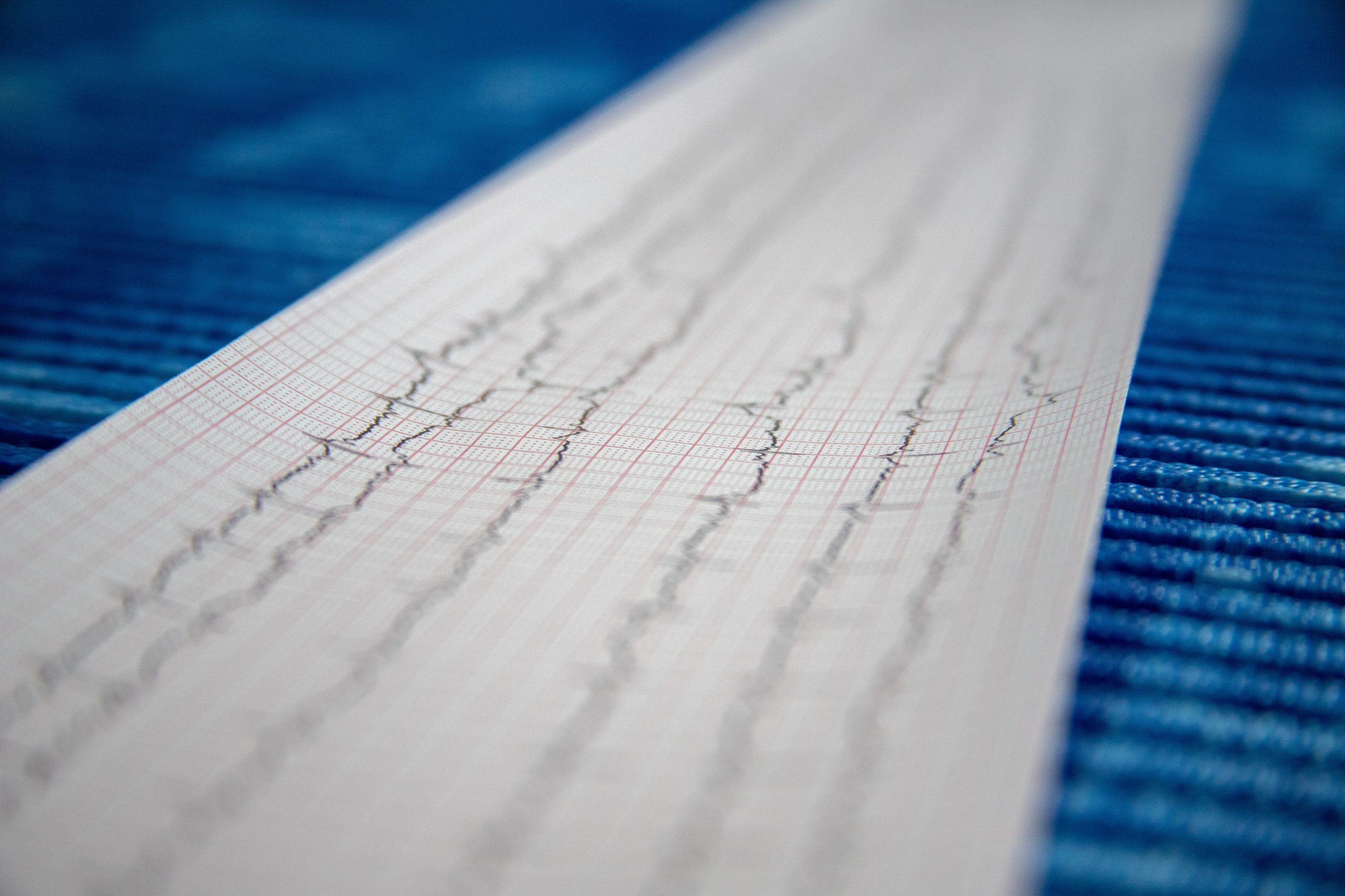
1. Stroke
Ischemic stroke is one the most common complication of afib that can cause mortality.
It constitutes around 80-90% of thromboembolic complications caused by afib.
Stroke can either be the first presentation of atrial fibrillation.
And there is a possibility that a patient already on blood thinners may still suffer from stroke.
Blood clots with afib are usually large in size and are formed in the left atrial appendage.
The size of a blood clot can range from a few millimeters to almost 4cm.
If afib remains for more than 48 hours, blood clots can be found in the atria of 5%-14% of patients.
These blood clots present in the left atrial appendage (LAA) can be visualized on transoesophageal echo (TOE).
They can dislodge from the heart and move to the blood vessels of the brain to disrupt the blood supply of that area.
When the brain cells remain deprived of blood above a certain time limit, they die & the function of that specific part of the brain is lost.
And this is what stroke is.
In the United States, afib has been shown to cause more than 70,000 ischemic strokes per year & this number contributed to 10-12% of total ischemic strokes.
The death rate after the first ischemic stroke within 30 days ranges from 16%-23%.
This means that out of every 100 people who suffer an ischemic stroke, 16-23 out of them die within the first month. (reference=UpToDate)
These mortality stats are for strokes without an underlying afib.
Studies have shown that when the stroke is secondary to afib, it is 2 times more fatal than without an underlying afib. [3]
How to recognise stroke in a patient with Afib?
In a patient of afib, the development of the following signs & symptoms is indicative of stroke:
- Sudden onset weakness which may involve arm, leg or a complete one half of the body.
- Sudden onset of inability to speak or understand speech.
- Inability to walk or loss of balance while walking which is also sudden in onset.
- Sudden onset of confusion or disorientation.
- Sudden onset of problems with vision.
A detailed physical examination along with the above-mentioned symptoms can confirm the diagnosis.
Imaging
CT scan
MRI
Management
Basic management principles for ischemic stroke are as follow:
- Thrombolysis in a subset of patients
- Mechanical thrombectomy
- Antiplatelets
- Lipid lowering drugs
- Blood pressure control
2. Acute Mesenteric Ischemia
Acute mesenteric ischemia is a condition that occurs when an artery (blood vessel) that supplies blood to the parts of the small intestine & large intestine gets occluded by the blood clot.
This can result in permanent damage of the affected area & can cause a widespread infection called sepsis & ultimately death.
The artery involved is called the superior mesenteric artery (SMA) & it delivers blood to the following areas:
- Small intestine including duodenum jejunum and ileum
- Appendix and cecum
- Large intestine (till 2/3rd of transverse colon)
The number of patients that develop acute mesenteric ischemia secondary to afib is low.
But the mortality rate i.e., the patients who die of this complication is quite high even after aggressive treatment.
Almost 50% of the patients with advanced ischemia requiring surgical intervention die of this complication.
How to recognise mesenteric ischemia in a patient with Afib?
Patients with acute mesenteric ischemia can have a widespread clinical presentation.
This makes it difficult to get recognized and is one of the factors that lead to a delay in treatment and increased mortality.
Some patients may remain completely asymptomatic.
Whereas some patients develop nonspecific symptoms like mild abdominal discomfort, nausea & fever, making it difficult to get diagnosed.
The typical presentation is as under:
- Severe abdominal pain which is sudden in onset & is way more in intensity than assessed by the physical examination.
This means that upon palpation of the abdomen there is no to very little tenderness, but the patient complains of severe pain.
- Nausea
- Vomiting
- Passage of fresh blood through anus
- Low blood pressure
Lab tests
Blood tests are non-specific.
They cannot establish a definitive diagnosis but can aid in the diagnosis.
These include the following:
- Raised white blood cells
- Raised hematocrit
- D-dimers
- Serum amylase levels
Plain X-rays
A plain x-ray of the abdomen again is a non-specific modality but still is widely used owing to its widespread availability.
It may show thickened bowel walls and dilated bowel loops.
Advanced imaging
These include CT angiography and MR angiography of abdominal vessels.
These procedures are just like CT scans or MRIs done for other purposes.
CT is more commonly done due to its lower cost and wide availability as compared to MR.
According to a study, the sensitivity of CT for diagnosing mesenteric ischemia is 93.3% whereas its specificity is 96%. [4]
This means that it can accurately establish a diagnosis of mesenteric ischemia in 93% of patients.
Management
Basic management steps include the following.
- Pain control which is usually done by opioids like morphine or nalbuphine.
- Blood thinners or anti-coagulants like heparin.
- Antibiotics for treatment of complications like sepsis.
- Intravenous fluids to maintain blood pressure or for management of shock.
- Abdominal surgery.
3. Acute Limb Ischemia
Blood clots formed in the heart can dislodge and travel to involve the blood vessels supplying arms or legs.
Sudden onset occlusion of blood supply to either arms or legs, caused by the blood clot is called acute limb ischemia.
As compared to stroke this complication of afib is less common.
Legs are more affected as compared to arms.
Atrial fibrillation remains one of the most important causes of acute limb ischemia.
Studies have shown that 60-95% undergoing surgery for embolism in peripheral circulation had underlying atrial fibrillation. (Reference: UpToDate)
Other causes include left ventricular clots secondary to a myocardial infarct or left ventricular dysfunction, infected or prosthetic heart valves throwing debris in peripheral blood vessels.
A high degree of morbidity and mortality is associated with acute limb ischemia.
Studies have shown that 30% of patients lose their limbs and almost 20% of the patients die. [5][6][7]
How to recognise acute limb ischemia in a patient with afib?
In contrast to mesenteric ischemia, acute limb ischemia presents with severe and typical symptoms.
This enables easy and early diagnosis.
The classic presentation is denoted by the famous 6Ps, which are.
- Pain (Severe acute onset)
- Pallor (an abnormally pale appearance)
- Pulselessness (absent pulses of the effected limb like absent radial pulse in case of arm involvement)
- Paraesthesia (burning sensation or a feeling of pins and needles)
- Paralysis (muscular weakness)
- Poikilothermia (cold limb)
- Another important symptom is cyanosis or bluish discoloration of involved limb.
Diagnosis
There are no specific lab tests.
Diagnosis is based on history, physical findings as mentioned above and imaging.
Imaging modalities include:
- duplex ultrasonography
- CT angiography
- MR angiography
- Catheter based arteriography which is used for diagnostic as well as therapeutic purpose as thrombolytic therapy can be instituted.
Management
Basic management steps are as under:
- Pain control
- Blood thinners
- Surgery
4. Heart Failure
Heart failure is another very important factor that contributes to mortality in patients with atrial fibrillation.
According to the European Society of Cardiology 2020 guidelines for atrial fibrillation, 20-30% of patients with afib develop heart failure & the mortality in such patients is significantly higher as compared to either of the condition alone.
Both these conditions often co-exist and are risk factors for each other
This means that the presence of atrial fibrillation can cause heart failure and the presence of heart failure predisposes a person for the development of atrial fibrillation.
In addition, the risk of clot formation is also increased when heart failure is present along with afib.
How Atrial fibrillation causes Heart Failure
AF can cause heart failure by a variety of mechanisms.
The details of those mechanisms are as under:
Increased heart rate and irregular rhythm make the contractions of the heart ineffective, thus reducing the cardiac output.
Persistently raised heart rate can cause a condition called tachycardia-induced cardiomyopathy which weakens the heart muscles and contributes to heart failure.
Increased concentration of certain hormones and chemicals that cause constriction in blood vessels like angiotensin II and nor-epinephrine.
How to recognise heart failure in a patient with Afib
History
Symptoms of heart failure and atrial fibrillation are usually overlapping.
Breathing difficulty, dizziness, & palpitations are the predominant symptoms.
One specific feature of heart failure could be breathing difficulty that worsens on lying straight called orthopnoea.
Physical examination
Physical examination may show the following signs:
Irregular low volume pulse
Low blood pressure
Cold peripheries
Presence of bilateral crackles in lungs on auscultation
Lab tests
Raised NT-Pro BNP levels can give a clue about heart failure, although it doesn’t confirm the diagnosis and co-relation with history, physical findings, & investigations is required.
However, normal levels of NT-Pro BNP exclude the presence of heart failure.
Imaging
Echocardiography
Cardiac MR
Diagnosis is made based on a combination of history, physical findings, lab tests, and imaging modalities.
Treatment
Basic management principles are as follows:
- Rate control with beta blockers or calcium channel blockers.
- Rhythm control with drugs like amiodarone or electrical cardioversion.
- Blood thinners either warfarin or NOACs like rivaroxaban.
- The drug of choice for heart failure with atrial fibrillation is digoxin.
- Diuretics
Take Home
Every patient with afib must be knowing all of the above complications as they can be fatal.
As soon as a patient develops any of these symptoms, he or she must immediately seek health care advice.
Adequate treatment for afib is the key for preventing these complications from happening.
Hi, I'm Dr. Junaid Arshad
Latest Posts
-

-

Can You Use Expired Glucometer Test Strips? What You Need to Know
November 24, 2021 -
-