Can Chest X-Ray Detect Heart Attack
-
- Dr. Junaid Arshad
- December 3, 2021
- 0 comments
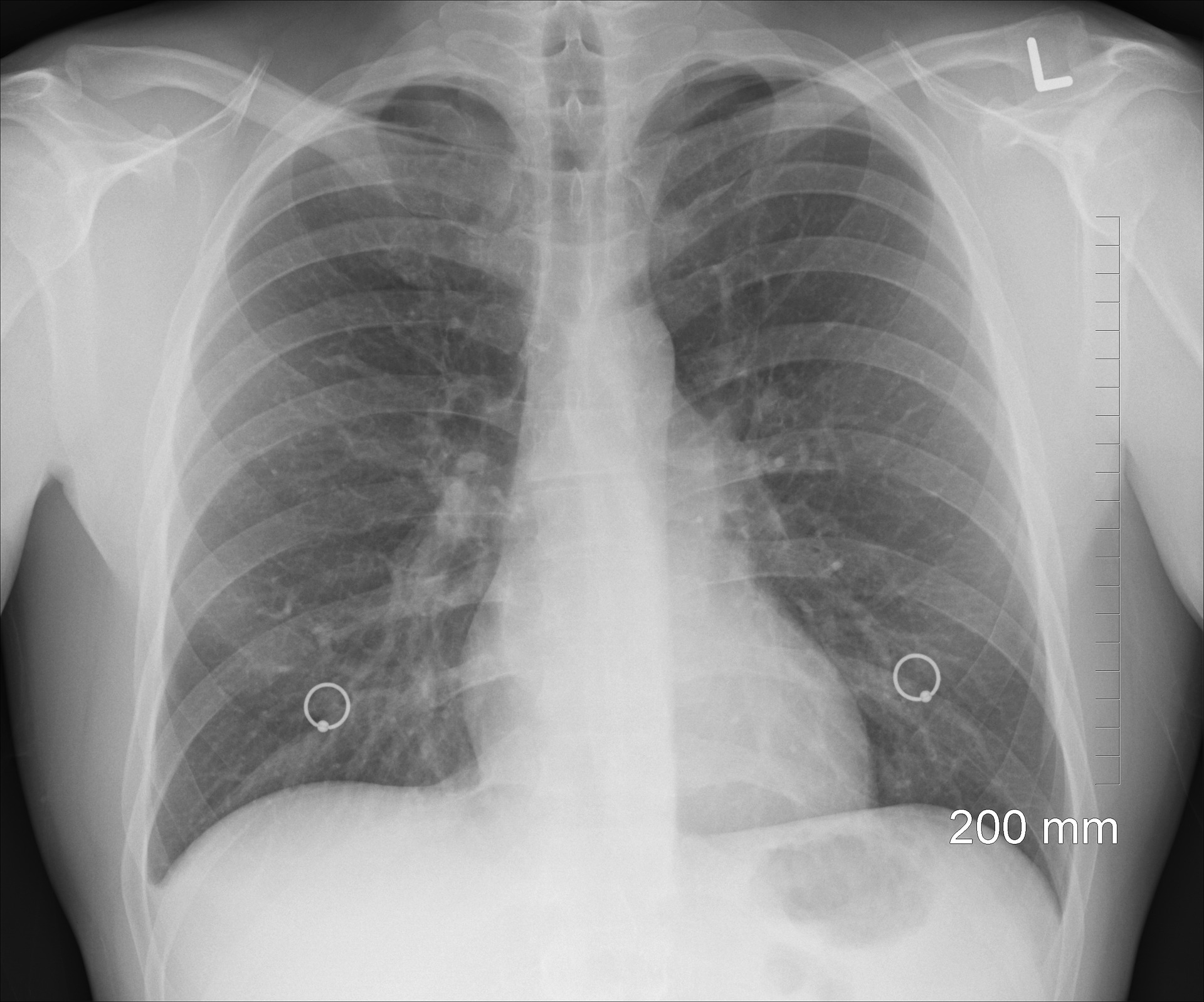
Chest X-ray (CXR) is the most commonly done imaging investigation in the entire world.
It is also the first-line imaging modality for the diseases affecting the lungs and the heart, & the other systemic diseases.
Table of Contents
Can Chest X-Ray Diagnose Heart Attack?
Answer
When it comes to heart attack, Chest X-Ray is not a very useful tool for establishing the diagnosis.
It cannot detect heart attack or acute myocardial infarct and is not done routinely for making the diagnosis.
However, it can detect a few complications that occur with a heart attack and are necessary to be diagnosed and managed.
Why CXR cannot detect Heart Attack
Answer
The reason it cannot diagnose a heart attack is that neither it can show regional wall motion abnormality like echocardiography nor it can show narrowing or blockage in the blood vessels of the heart like angiography.
Explaination
During a heart attack, what happens is, that the blood vessels supplying the heart muscles get clogged by the cholesterol depositions.
This results in a complete cessation of blood supply to that particular region of the heart supplied by the affected vessel.
When heart muscles remain deprived of blood above a certain time period, they stop functioning and ultimately get permanently damaged.
The entire process ends up in the following 4 consequences.
#1. Decrease blood supply to the heart, affects the electrical activity required for normal contraction of heart muscles. This change in electrical activity is detected by the EKG making it the 1st and the most important initial investigation.
#2. Heart muscle damage causes an enzyme present inside the myocardial cells to leak and get detected in the blood. It is called troponin and its presence in blood confirms the diagnosis of heart attack.
#3. The region of heart having compromised blood supply stops functioning. In other words its contractility reduces & this can be seen on echocardiography. This is how echo can help in the diagnosis.
#4. Blocked blood vessel can be seen on angiography which is the gold standard for the diagnosis and guides further management as well.
Unfortunately, Chest-Xray cannot detect any of the above 4 abnormalities.
Hence in acute MI, it has a limited role only in the detection of a few complications mentioned below.
3 Abnormalities caused by Acute MI (Heart Attack) which Chest X-Ray can detect
Chest X-Ray is normal in up to 50% of the patients presenting with a heart attack.
It cannot detect ongoing heart attacks but as previously mentioned can help identify a few complications.
These include the following:
#1. Pulmonary Edema
It is a condition in which excessive fluid accumulates within the lungs and hence patient experiences breathing difficulty.
Breathing difficulty can sometimes be severe enough to require mechanical ventilation & can be life-threatening.
CXR is the most practical modality for the detection of pulmonary edema.
It can also quantify heart size and help identify if pulmonary edema has recently developed after a heart attack or if it’s present due to a previously ongoing disease like cardiomyopathy.
In case of pulmonary edema with a recent MI (Heart attack), heart size is normal.
On the contrary, when pulmonary edema is secondary to chronic heart failure or any other cardiomyopathy, the heart size is usually enlarged called cardiomegaly.
Mechanisms of pulmonary edema with Heart Attack
There are 2 mechanisms that lead to pulmonary edema after a heart attack.
- Heart attack affects a specific region of heart muscle depending upon the blood vessel involved.
Sometimes the affected region is large enough to decrease the pumping ability of the heart.
This causes back pooling of blood and in turn an increased pressure within the blood vessels of the lungs which ultimately causes pulmonary edema.
2. The second mechanism is a mechanical defect called MR (mitral regurgitation) which in common terms is called valve leakage.
It occurs due to rupture of the papillary muscle & the rupture itself occurs due to reduced blood supply.
Papillary muscles are the muscle projections that are attached to the valve and keep the valve in place.
As the mitral valve gets leaky, there is a backflow of blood in the left atrium and the blood vessels of the lungs which cause pulmonary edema.
Clinical Importance of CXR in Heart Attack
So CXR when done in patients with a heart attack can diagnose this life-threatening pulmonary edema and give a clue that heart attack has involved a large area of the heart & this has prognostic importance.
Patients who develop heart failure with heart attack have worse outcomes as compared to those who don’t.
#2. Ventricular Aneurysm
This is one of the worst complications that can occur after a heart attack and is visible on CXR.
An aneurysm is a term that refers to a bulge or an outpouching that forms in weakened heart muscles.
This complication can cause heart failure and life-threatening rhythm abnormalities and hence can contribute to mortality. [1]
Both anteroposterior and lateral views can show the presence of an aneurysm.
#3. Pericardial Effusion
CXR can show the pericardial effusion which is the presence of fluid in the cavity around the heart.
Dressler’s syndrome, one of the delayed complications of a heart attack occurring after 4-6 weeks can cause pericardial effusion.
Alternate of Chest X-Ray for detecting Heart Attack
The alternate imaging modality that has a diagnostic role for heart attack is echocardiography.
It is easily available in cardiac setups, is inexpensive, and is a noninvasive investigation that can yield valuable information.
As previously mentioned, during a heart attack the contractility of the affected muscle is reduced and can be seen on echo.
In fact, echo can detect this decreased contractility as soon as the blood supply to heart muscles is decreased and even when EKG changes have not been developed.
This makes EKG a global recommendation by the authorities like European Society of Cardiology (ESC) for the diagnosis of a heart attack.
What other heart abnormalities with Coronary Artery Disease can CXR detect?
- Coronary Artery Calcifications
- “Coronary artery calcification triangle” is a site on CXR which can show blood vessels of heart & includes the left main, LAD & LCx.
- Lateral chest x-ray can show another vessel called RCA.
- Calcified coronary artery aneurysms.
Summary
Chest X-Ray has got no role to diagnose an Acute Myocardial infarction but it can show a few complications like pulmonary edema which refers to excessive fluid in the lungs.
An alternate imaging investigation for the diagnosis of MI is echocardiography which can detect the decreased contractility in the affected region of the heart.
Hi, I'm Dr. Junaid Arshad
Latest Posts
-

-

Can You Use Expired Glucometer Test Strips? What You Need to Know
December 3, 2021 -

-