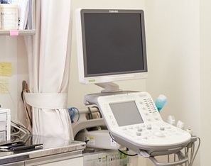
Can You have heart disease with a normal echocardiogram?
-
- Dr. Junaid Arshad
- March 26, 2022
- 0 comments
Yes, a person can still have heart disease even if the echocardiogram is normal. Echo can detect both structural and functional abnormalities of the heart but diseases like Coronary Artery Disease(CAD) may not produce any significant defect in both the structure and the function until it causes a heart attack & damages the heart muscle.
A damaged heart muscle can be seen on echo in the form of decreased contractility of the affected segment. But a significant CAD characterized by severe narrowing in the blood vessels of the heart (coronary arteries) may not result in a heart attack and in such cases echo may show a completely normal heart.
Echo cannot visualize the coronary arteries and can be normal even with significant narrowing in coronaries. This narrowing can be seen on angiography.
Explaination
To have a better understanding of the subject, let us review the normal functioning and blood supply of the heart.
The heart is an organ that pumps blood to all the organs of the body.
It is made of 4 chambers. The upper 2 chambers are called atria and the lower 2 chambers are called ventricles.
Both atria and ventricles are made up of heart muscle called the myocardium.
Just like any other organ, the heart itself needs oxygen to remain healthy and carry out its function. This is supplied through the blood.
The heart receives blood from the following 3 major vessels.
- Left anterior descending artery (LAD)
- Left circumflex artery (LCx)
- Right coronary artery (RCA)
Also, there are microvessels that supply the heart muscle.
In the case of CAD, the cholesterol gets deposited in the blood vessels and causes them to get narrowed over time.
The narrowing in coronary vessels decreases the blood supply of heart muscle.
A patient may remain completely asymptomatic with as much as 70-80% narrowing in coronary vessels. Whereas, some patients may experience anginal pain. However, a resting echocardiogram is usually normal in both of these.
Role of stress Echocardiography
A solution to the above-mentioned problem is stress echocardiography.
Stress echo is just like a normal echo but a patient’s heart is put under stress.
This is done by giving a medicine called dobutamine which is given intravenously.
Dobutamine acts on the heart to increase both the force of contraction & the heart rate.
This increases the workload of the heart and in turn the oxygen requirement of the heart muscle.
If there is narrowing in the coronary vessels, the involved segment of the heart would not receive the required blood & this can be seen on the echo in the form of decreased contractility.
This abnormality cannot be seen on resting echo, which may show a completely normal heart.
This raises another question, does every patient with a normal echo should undergo a stress echo?
The answer is ‘No’.
Stress echo is required to have a clue of CAD only in patients with a very high probability of CAD.
The high probability of CAD is indicated by the history of typical anginal pain & the presence of risk factors like family history, diabetes, hypertension, smoking, obesity, etc.
Other Heart diseases that do not show up on the Echo
Other diseases that may give a completely normal echo are given below.
Microvascular angina
Microvascular angina(MVA) aka Cardiac Syndrome X causes narrowing in the small vessels of the heart.
The patients experience typical anginal pain.
But as it involves only the microvasculature, the diagnosis is difficult to establish.
Resting ECG and echo both are normal.
Also, the angiogram shows normal unobstructed coronary vessels or a narrowing of 50% or less.
As mentioned previously for CAD, not every patient with chest pain and normal investigations should be considered for MVA. This should be decided by the cardiologist, based on the patient’s history and clinical suspicion.
Rhythm Abnormalities
Rhythm abnormalities of the heart like atrial fibrillation, supraventricular tachycardias, & WPW syndrome may also show a completely normal echo.
But sometimes they can be due to structural abnormalities which can be seen on echo for e.g mitral valve disease in case of atrial fibrillation.
Which disease echocardiogram can detect?
Echocardiography is one of the most commonly used diagnostic modalities in cardiology.
As compared to other diagnostic modalities, it is cheaper, easily available, noninvasive, & can quickly give comprehensive information about both the structure and function of the heart.
The following heart diseases can be detected on echo:
Ischemic Heart disease
In the case of heart attacks echo is one the of key diagnostic modalities.
Estimation of the ejection fraction by the echo guides further therapy.
It helps u determine which segment of the heart is affected by the heart attack.
The affected segment is determined by the reduced contractility termed hypokinesia or akinesia.
Mechanical complications that occur after a heart attack like mitral regurgitation(MR), ventricular septal rupture(VSR), & free wall rupture are also diagnosed on echo.
Valvular heart diseases
Echocardiography is the investigation of choice for establishing the diagnosis and severity of valvular diseases like mitral stenosis, mitral regurgitation, aortic stenosis, aortic regurgitation, & also for pathologies of pulmonary & tricuspid valves.
Determining the type of valvular lesion and its severity helps guide therapy like medical management of surgery like valve replacement.
Pericardial effusion
Pericardial effusion means the presence of excessive fluid within the pericardium.
The pericardium is a sac-like double-layered membrane that surrounds the heart.
Excessive fluid in the pericardial cavity can affect the functioning of the heart resulting in a condition called cardiac tamponade, which is a cardiac emergency and requires immediate treatment.
It is diagnosed clinically by the presence of a triad of signs called beck’s triad which includes low blood pressure, engorged neck veins, & muffled heart sounds.
The condition is confirmed through echocardiography which is both sensitive and specific for establishing the diagnosis.
Cardiomyopathies
Cardiomyopathies are disorders affecting the heart muscle.
In these disorders, the heart is both structurally and functionally abnormal.
Following diseases are classified as cardiomyopathies:
● Dilated cardiomyopathy (DCM)
● Hypertrophic cardiomyopathy (HCM)
● Restrictive cardiomyopathy (RCM)
● Arrhythmogenic right ventricular cardiomyopathy/dysplasia (ARVC/D)
Echocardiography is the initial and one of the important investigations for all sorts of cardiomyopathies.
The details of echocardiographic findings are beyond the scope of this article.
Myocarditis
Myocarditis refers to inflammation of the heart muscle.
It can be due to infectious or noninfectious causes.
Echocardiography is the key initial diagnostic investigation.
LV dysfunction and dilated LV are the common findings.
Wall motion abnormality is usually global as compared to segmental in the case of ischemia.
Confirmation of myocarditis is done on cardiac MRI.
Pulmonary embolism
Pulmonary embolism is a condition characterized by occlusion of the pulmonary artery or its branches by the blood clot.
It is an emergency situation and can be fatal.
It is a fairly common condition.
In the US it accounts for 100,000 deaths in a year.
In Europe, pulmonary embolism causes around 300,000 death every year.
Men are more affected than women with an estimated mortality of 56 versus 48 per 100,000, respectively.
It is one of the most missed diagnoses.
Echocardiography plays a supportive role in the diagnosis of pulmonary embolism.
Right heart strain and dilated right chambers can be seen on echo. Sometimes a clot can also be seen in the right heart & rarely in pulmonary arteries.
Other echo findings include decreased RV function, tricuspid regurgitation, abnormal septal wall motion, McConnell’s sign
CTPA confirms the diagnosis.
Clots/Masses
Blood clots or tumors can be easily diagnosed on echo.
Cardiac MRI or CT scan may be required in some cases.
Congenital heart diseases
Congenital heart diseases can be diagnosed via echocardiography as it gives information about both the anatomy and functioning of the heart.
Common ones include the following:
Atrial Septal defect
Ventricular septal defect
Coarctation of aorta
Patent ductus arteriosus
Ebstein’s anomaly
Tetralogy of Fallot
Transposition of great arteries
Truncus arteriosus
Categories
- Cardiology 32 Posts
- Diabetes 4 Posts
- Drugs 1 Post
- Infectious Diseases 2 Posts
- Sexual Health 3 Posts


